
Abstract
Introduction
Large impaled object in the orbital region causes severe visual impact and requires specialized care within the shortest time possible.
Objectives
In this case report, we discussed the approach and management of a patient that presented with a penetrating orbitocranial injury, from management at the emergency unit, diagnostic imaging, referral to other subspecialty, surgical and medical intervention, and post-operative care.
Discussion
A 36-year old male had an impaled toilet brush on the supero-nasal aspect of the right orbit, with visual acuity of 6/60 and lacerated upper eyelid. The globe had minimal movement on all gazes, but pupil was reactive to light with no afferent defect. On plain cranial and orbital CT-scan, the foreign body entered the anterior and medial aspects of the right orbit penetrating the right superior orbital wall, right medial lamina papyracea, and the lateral and inferior border of the right frontal sinus with its distal tip at the intracranial region at the right frontal lobe compressing the medial rectus along its tract. Two hours after injury, patient underwent wound exploration, removal of foreign body, repair of eyelid laceration, right craniotomy, frontal contussectomy, duraplasty, and JP-drain insertion under general anesthesia. Intraoperatively, there was note of transected canaliculus and avulsed conjunctiva. The medial rectus was intact and attached. The frontal lobe was contused with embedded fragments of right posterior orbital bone with 3cm opening on the dura. Post-operatively, Fluconazole was added to the medications after culture results of the toilet brush tip tested positive for fungal elements. Patient was discharged after 21 days with visual acuity of 6/6 on both eyes and improved ocular movement.
Conclusion
These types of injury warrants thorough and systematic history taking and physical examination, acquiring pertinent imaging modalities to better visualize the extent of injury, and execute surgical and medical intervention that is multidisciplinary.
Author Contributions
Academic Editor: Asaad Ghanem, Mansoura ophthalmic center, mansoura university, mansouraam, Egypt.
Checked for plagiarism: Yes
Review by: Single-blind
Copyright © 2021 David G. Diciano, et al.
 This is an open-access article distributed under the terms of the Creative Commons Attribution License, which permits unrestricted use, distribution, and reproduction in any medium, provided the original author and source are credited.
Competing interests
The authors have declared that no competing interests exist.
Citation:
Introduction
Large impaled object in the orbital region causes severe visual impact and requires specialized care within the shortest time possible. The urgency of the injury is brought about by the different organs that are possibly affected, and may lead to severe debilitating complications.
Objectives
The Objectives of this Case Report are as Follows:
To present a case, and discuss the management of an orbitocranial penetrating injury seen at the RMC-Emergency Department
To discuss the appropriate approach to patients with orbitocranial penetrating injury.
Identifying Data
A 36 year old male from Sto.Tomas Pasig, Philippines, presented at the emergency room with an impaled toilet brush to his right eye. Approxiamtely 35 minutes from the consults, patient woke up and went to relieve himself. Inside the comfort room, he slipped, falling face first hitting a toilet brush with his right eye. Patient was immediately brought to Rizal Medical Center. There was no note of loss of consciousness and vomiting. Patient is allegedly not drunk.
At the emergency room, patient was on a wheel-chair, oriented to three spheres, cooperative, and with minimal amount of pain. Patient was slightly tachycardic but was adjudged with stable vital signs.
On reading distance the visual acuity of the right eye was J20 while it was J1 for the left eye.
Grossly, for the right eye, there is a penetrating foreign body on the superomedial aspect of the orbit (probably extending to the inner medial wall). The upper lid is transected with minimal soft tissue swelling. There is a sero-sanguinous discharge on the site of insertion but no active bleeding was noted. The globe seems intact, and the pupils were 2-3mm briskly reactive to light. No RAPD was noted.
The left eye is grossly normal, with pupils 2-3mm briskly reactive to light with no relative afferent pupillary defect. Figure 1.
Figure 1.images 1-3 are that of the patient upon examination at the emergency unit.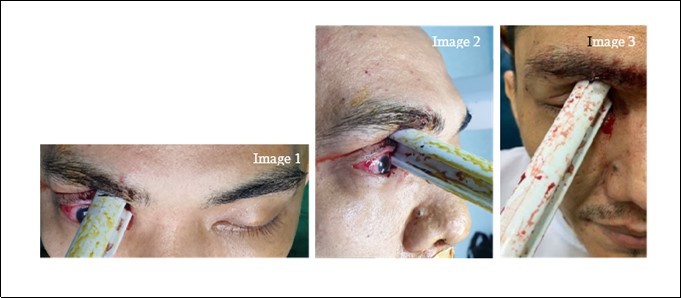
Upon examining the extraocular muscles, the right eye seems to be frozen with minimal movement on vertical gazes and no movement at all on horizontal gazes. The left eye however exhibits full muscle activity. Digital palpation was deferred.
On direct funduscopy, the right eye exhibits red-orange reflex. We were unable to go further with this examination for the right eye because any maneuver nearer the right eye was inhibited by the length of the toilet brush. The left eye however, exhibited red orange reflex and clear media. The cup-to-disc ratio is 0.3 with distinct disc boarders. The arterio-venous ratio was 2:3, non-tortuous, and follows a normal course. There was good foveal reflex. There were no hemorrhages and exudates.
The rest of the neurologic examination was normal.
At this point, the primary working impression was orbital penetrating injury, on the right eye with an impaled foreign body, upper eyelid margin laceration, with possible transection of the upper canaliculus, and to rule out globe pathology.
Patient underwent emergency cranial CT SCAN with orbital cuts to evaluate the extent of the injury. Figure 2.
Figure 2.The images (4-6) on the upper row, are serial coronal cuts of the cranial CT scan done for the patient. While images on the lower row (7-9) are axial cuts of the same scan.
The images (4-6) on the upper row, are serial coronal cuts of the cranial CT scan done for the patient. While images on the lower row (7-9) are axial cuts of the same scan.
There is a barrel shaped hypodense foreign body, seen to enter the anterior and medial aspects of the right orbit. The aforementioned foreign body is directed superiorly and is seen traversing or penetrating the right superior orbital wall, right medial lamina papyracea and the lateral and inferior border of the right frontal sinus, with its distal tip at the intracranial region at the right frontal lobe. There are fractured fragments, likely from the comminuted superior orbital wall and seen within the right frontal lobe. Along its track, it compresses the right globe and the right medial rectus muscle. Pneumocephalus in the right frontal convexity was noted and hemosinus in the right frontal and ethmoid sinus are likewise observed. Prolapse of the right medial orbital fat into the ipsilateral ethmoid sinus is noted.
With the results from the cranial CT scan, the patient was assessed to have orbitocranial penetrating injury on the right eye, with an impaled orbitocranial foreign body (plastic), an upper eyelid laceration with possible transection of the upper canaliculus, multiple orbitocranial bone fractures (Right frontal, ethmoid, sphenoid bones), and presence of pneumocephaly (frontal area). At this point we still can not totally rule out globe pathology.4, 7
The patient was referred to the neurosurgery service that took over as the primary service provider of the patient.
The combined surgical plan was emergency wound exploration, removal of foreign body, repair of eyelid laceration, of the right eye care of ophthalmology service followed by right craniotomy, frontal contussectomy, duraplasty, and JP drain insertion c/o Neurosurgery service all under general anesthesia.
Patient was roomed to the operating room, 2 hours post-injury. While preparing the field of operation the foreign body was held into a fixed position and even while the patient was being intubated by the anesthesiologist. Another resident prepared and sterilized the field of operation. The brush part of the foreign body was covered with sterile gloves while the handle was generously washed with betadine solution.
Emergency would exploration was done. After careful examination of the extent of the wound, the foreign body was mobile enough to be carefully lifted.
Intraoperatively, there was note of transected canaliculus and avulsed conjunctiva. The medial rectus is intact and attached. Bone fragments were also recovered from the wound site. It was also noted that the frontal lobe was contused with embedded fragments of posterior orbital bone and 3cm opening of the dura. The tip of the brush that was embedded to the eye was sent for culture examinations.
On day 1 post-operation, patient was awake, comfortable, not in pain, and was oriented to 3 spheres. On reading distance, the visual acuity of the right eye was J5 and the left eye was J1. Patient was able to perfectly identify all 15 plates of the Ishihara test. Grossly on the right eye, the sutures were in place. No wound dehiscence noted nor was there any serosanginous or foul-smelling discharge. Pupils for both eyes were 3-4mm briskly reactive to light with no note of relative pupillary defect.
Upon testing for the extraocular muscles, the right eye exhibits, (-4) limitation on vertical gazes, (-2) adduction deficit, and (-4) abduction deficit. Patient noted diplopia on all extreme gazes, however there was no pain. The left eye exhibited full activity of its muscles.
On funduscopy both eyes exhibited red-orange reflex, clear media, distinct optic disc boarders with a cup-disc-ratio of 0.3. The arterio-venous ratio was 2:3, following a non-tortuous normal course. There were no exudates and hemorrhages noted. There was good foveal reflex for both eyes.
On repeat CT Scan, day 1 post-operation, a craniotomy defect was seen in the right frontal bone involving the right frontal sinus. The previously noted foreign body entering the right orbit was no longer seen. The right medial rectus muscle remains thickened with lessened medial prolapse into the ipsilateral ethmoid sinus. The right inferior rectus was also slightly thickened when compared to the left. Minimal fat stranding was seen in the right intraconal space. The right globe appeared intact. The comminuted fractures of the right medial orbital and ethmoid sinus remains seen. Fluid densities with hyperdensities were still seen in the right ethmoid sinus and with slight increase.
Course in the Wards:
Post-operative day 1, the ophthalmology service suggested starting the patient on steroids pending the results of the culture examination of the toilet brush tip, and approval from co-managing services.
Post-operative day 2, aside from the initial antibiotic given, the primary service included Vancomycin upon the prescription of the Infectious Diseases-service.
Post-operative day 4, culture results tested positive for fungal elements. Vancomycin was discontinued and patient was shifted to Fluconazole 400mg IV for 14 days.
Post-operative day 13, from (-3) adduction deficit of the right eye, it improved to (-2). The visual acuity was also improved from J5 to J3.
Post-operative day 16, the visual acuity of the right eye is now J1.
Post-operative day 18, visual acuity of the right eye improved to 6/6. Limitations of the gazes were also improved.
On day 21, patient was discharged. Sutures on the lids were already released, and there was no note of wound dehiscence. We also noted a decrease in palpebral fissure of the right eye compared to the left which could be due to remnant inflammation. There was no complaint of epiphora from the patient. We also noted a significant improvement in vertical gazes and minimal improvement on horizontal gazes.
Discussion
Penetrating eye injuries to patients occur most commonly on the 20-40 years age group, the supposedly most productive workforce of the population, with males more commonly involved than females. Hammering metal objects was the most common mechanism of injury1.
When presented with patients having orbitocranial penetrating injury, we are reminded to (1)protect the eye as much as possible to limit and avoid manipulation that might worsen the extent of injury. (2)Use shields (plastic shield applied with transpore tape). If not available we improvise using cut-down polystyrene drinks cup. We ensure it is securely attached but also cautious not to exert any pressure to the eye. (3) Keep patient nothing by mouth in anticipation of possible surgical intervention under general anesthesia. When we think about analgesia, there are varieties of routes. It could be topical/IM/IV. Note that topical anaesthetics should be used sparingly as it carry a risk of corneal toxicity. (4)Consider antiemetic. (5) Check tetanus status and give booster if necessary1.
It is also important not to (1)Try to remove embedded foreign bodies. (2) Apply eye pads. (3) Put pressure that can cause herniation of ocular contents. (4)Try to clean the eye as we may inadvertently remove ocular contents (e.g. uvea or retina). (5) Put cream or ointments if we are not sure that the globe is intact. The creams and ointment might sieve through a perforation and be toxic to the retina. (6) Delay referrals1.
Factual details are essential in determining the type of injury and enable rapid stratification of likely risk to the eye. Important factors to asks include: When?: Timing of injury (and any delay to presentation with reasons). Where?:Occupational or assault-related injuries should be carefully documented for medico-legal reasons. How?: Determines the trajectory of the object and give an idea of the extent of the injury. Description of object causing injury, size, weight, velocity, direction (head-on or glancing impact)1.
When examining the eye, it is imperative that the patient is stable both haemodynamically and neurologically1.
A drop of topical anaesthetic is a great aid to examination. Eyelids are very infrequently ‘‘too painful to open’’. Eye drops containing preservatives are best avoided as allergy can occur and they are retinotoxic if a penetrating injury is present1.
Eyelids should never be forced apart. If topical anaesthesia and verbal encouragement are not sufficient then examination by a clinician with experience of ocular trauma is usually indicated. If the history is suspicious of penetration then this alone should require early specialist ophthalmic input1.
For cases with legal implications, we document properly our findings and use pictures if necessary and if the law allows1.
When examining the eye, it is important to do it in a systematic manner. Pupillary reactions are assessed first followed by, visual acuity, and then ocular motility. When assessing the extent of injury, we do it front-to-back and then compare with the other eye (especially if the other eye is not affected). We first check for the lids, then the anterior segment. The anterior segment is best examined under a slit lamp; if not available or limited by the extent of injury, a portable slit lamp makes a good alternative. Then the posterior segment, ideally examined with the use of mydriatics1.
Investigations
Plain facial X-rays are used for the detection of orbital fractures, however, their role in some cases of suspected intraocular foreign body is questioned.2,3Note should be made that radiolucent material is not visible on plain films and again, the history should raise your suspicion of intraocular foreign body due to the mechanism of injury1,3.
The orbit is very fragile and susceptible to injury. Double vision and restricted eye movements would direct you to a possible orbital fracture. These can be apparent on plain films but other features such as fluid levels in sinuses can be the only sign. CT again is the gold standard3.
CT-scan of the orbits is useful for imaging the orbit in greater detail as bony injuries are more apparent. The globe is well defined and perforations can be detected more easily. It can also be used for detection of intraocular foreign bodies and globe contents within the orbit indicating perforation1, 4.
(Figure 3) is a prescribed simplified approach in investigating and managing patients with intraocular penetrating injuries. If we are going to use a non-contrast CT, we ask. If we suspect vascular injury, cranial nerve injury, or fractured of sphenoid wing. To such cases an CT Angio or MRA is encouraged. If not, we ask if the foreign body is wood. On such cases, and MRI is better. If not then we proceed on giving antibiotics and consider giving steroid, whenever safe, to protect the nerve. If there is CSF leak, displaced fracture, ICH, or vascular injury, then we operate immediately. If not, we ask if the foreign body is still there. If yes, then we do removal under general anesthesia. If the foreign body is no longer on the wound site, then conservative management is sometimes warranted more5,4.
Figure 3.Flowchart adopted from Screckinger, 2011 on how to choose the proper diagnostic imaging depending on the injury.
Treatment
It is recommended to initiate treatment as soon as admission. Broad spectrum with good CNS penetration is generally advised (Ceftriaxone, Ciprofloxacin, Metronidazole) We also send the foreign body for culture (specially if wood)5,6,7.
High dose steroids indicated for traumatic optic neuropathy but visual outcome benefits are yet to be proven5,6.
The decision to operate on a patient with penetrating transorbital injury should be made as a collaborative effort between members of the neurosurgery, ophthalmology, otolaryngology, and maxillofacial surgery teams5.
Typically, the role of the neurosurgeon is to decompress the brain and neurovascular structures, prevent injury to intracranial structures during removal, and assist in the reconstruction of the skull base. Subsequently the expertise of otolaryngologists and maxillofacial surgeons is needed to stabilize the orbital, maxillofacial, and rhinological structures5,6.
Indications for surgery includes retained foreign body, presence of dural defects (especially with CSF leakage immediately after trauma), displaced bone fractures, intracranial hematoma, and evidence of direct vascular injury.Some authors have suggested that emergency surgery is indicated in unstable patients with extended transorbital brain injuries in lieu of cerebral angiography to allow for prompt decompression and hemostasis5,7.
The initial goals of surgery are decompression of neurovascular structures and removal of the foreign body under direct visualization. After the foreign body is removed, the operative goals shift to removal of bone fragments, repair of fractures, debridement of involved parenchyma, hemostasis, and dural closure. After surgical repair of intracranial injuries, the globe is evaluated extensively for injury.Postoperatively, CT scanning or MR imaging must be performed to rule out the presence of new/missed hematoma or retained foreign body5,6,7.
Complications
There are a number of possible complications after transorbital, intracranial penetration of a foreign body. The transorbital penetration itself can cause specific complications related to the path by which the foreign body exits the orbit. When the object passes through the superior orbital fissure, it can cause orbital apex syndrome, which involves injury to the oculomotor nerve (CN III), trochlear nerve (CN IV), abducens nerve (CN VI), ophthalmic branch of the trigeminal nerve (CN V1), and the optic nerve (CN II), resulting in loss of vision and opthalmoplegia5,6.
If the object penetrates the cavernous sinus, it can cause cavernous sinus syndrome, which can manifest similarly to orbital apex syndrome, with the addition of injury to the maxillary branch of the trigeminal nerve (CN V2) as well as the oculosympathetic fibers, resulting in facial numbness and miosis5,7.
Penetration through the orbital roof often results in frontal lobe contusions. A CSF leak can also occur due to the trauma, but if one is not initially evident, we do not recommend operating prophylactically to try to prevent one from developing. Prophylactic operation exposes the patient to the risks inherent to surgery, and a CSF leak can usually be managed conservatively5,7.
Trauma to the globe can result in sympathetic ophthalmia, an autoimmune process resulting in bilateral, nonnecrotizing granulomatous uveitis. While the exact pathophysiology is not understood, it is believed that an autoimmune reaction develops in response to exposed ocular antigens after globe disruption. Since the development of sympathetic ophthalmia is a rare event and is delayed by up to 10 days following globe injury, enucleation can be performed at a separate sitting, allowing acute neurosurgical issues to be treated first5,6,7.
Post-operative complications after repair of a penetrating orbital injury include CSF leak, meningitis, cerebral abscess, carotid cavernous fistula, traumatic aneurysm, and progressive intravascular thrombosis. When a patient develops a cerebral abscess postoperatively, a retained foreign body should be ruled out5.
Conclusions
1. Large impaled objects in the orbital region can cause severe visual impact and require specialized care within as short of a time as possible.
2. Careful, thorough, and systematic history taking and physical examination is imperative
3. Investigations with imaging modalities is necessary to better visualize the extent of injury.
4. Approach to these types of injuries should be sequential and multidisciplinary.
References
- 1.Lindfield D, Das-Bhaumik R. (2009) Emergency department management of penetrating eye injuries. , International Emergency Nursing 17, 155-160.
- 2.Saeed A, Cassidy L, D E Malone, Beatty S. (2008) Plain X-ray and computed tomography of the orbit in cases and suspected cases of intraocular foreign body. , Eye 22(11), 1373-1377.
- 3.Salvolini U. (2002) Traumatic injuries: imaging of facial injuries. , Eur. Radiol 12(6), 1253-1261.
- 4.Fezza J, Wesley R. (1999) Importance of CT Scans in Planning the Removal of Orbito- Frontal Lobe Foreign Bodies. Ophthalmic Plastic Surg. 5(5).
- 5.Screckinger M.Transorbital penetrating injury: case series, review of the literature, and proposed, management algorithm. , J Neurosurg 114, 53-61.