
Abstract
Reaching efficient, safe and painless medical diagnosis procedure is a very valued goal for many research areas. Despite the great advantages of using optical imaging techniques in medical diagnosis including high safety and relative simplicity, it still suffers from relatively low resolution and penetration depth in the multiple scattering mediums such as biological tissues. Therefore, researchers began to devise ways to reduce the scattering properties of the tissue, hence increasing the imaging contrast. Optical clearing concept is introduced to do this job. This technique can reduce tissues scattering properties by using high refractive index chemicals, thus making the tissue transparent by equalizing the refractive index through that medium. In this paper, theory and techniques of optical clearing method are illustrated utilizing its benefits for deep imaging of different body tissues and organs.
Author Contributions
Academic Editor: Angela Pia Cazzolla, University of Bari, Italy.
Checked for plagiarism: Yes
Review by: Single-blind
Copyright © 2020 Omnia Hamdy, et al.
 This is an open-access article distributed under the terms of the Creative Commons Attribution License, which permits unrestricted use, distribution, and reproduction in any medium, provided the original author and source are credited.
Competing interests
The authors have declared that no competing interests exist.
Citation:
Introduction
The relatively low optical penetration as a result of the scattering properties of biological tissues is considered the main limitation of many optical imaging techniques. The principal reason of this multiple scattering characteristics is the difference in the refractive indices between the tissue cell components and its background 1. The tissue components such as cell nuclei, collagen, elastin fiber, mitochondria, organelles, cytoplasm and plasma membrane of the cell have refractive indices ranging from 1.47 to 1.51, while the surrounding medium has refractive index equals to 1.33, similar to that of water. This causes light diffusion and scattering inside biological tissues 2. Therefore, a trend has emerged from scientific research towards discovering ways of reducing scattering basically by means of refractive index matching. This research area has been named optical clearing 3. From the beginning of the 21th century, much more research items related to optical clearing techniques started to show up with a great increasing from 2013 till now 4. The method started to attract researchers from different areas including physics, chemistry, bioengineering and medicine. Recent studies have been aimed at altering turbid biological tissues in such a way that they become more optically transparent while keeping their internal structure intact using chemical compound with distinct osmotic properties such as glucose and glycerol 5.
After applying optical clearing agents (OCAs), tissue optical scattering parameters “anisotropy and scattering coefficient” have to be calculated to ensure the reduction in the scattering properties of the treated tissue. Determination of tissue optical parameters can be achieved using either integrating sphere measurements 6 or distant detector based configuration 7.
Optical Clearing Techniques
Tissue optical clearing can be achieved using different techniques; physical, chemical, photo-chemical/photo-thermal and compression. However, choosing the suitable clearing protocol is mostly depending on the size and the physical properties of the analyzed tissue sample 8. The schematic diagram presented in Figure 1 summarizes the main clearing techniques.
Figure 1.Different clearing techniques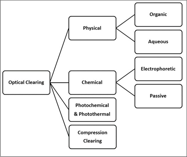
In the physical optical clearing approach, the clearing is achieved by using refractive index matching solutions in order to reduce the optical inhomogeneity of the tissue sample, these solutions can be either organic or aqueous 4, 9. The organic optical clearing agents are organic solutions with high refractive index and the clearing process is implemented by two steps: dehydration and clearing. In the dehydration step, the water content of the studied sample is removed commonly using methanol, ethanol, tert-butanol, or tetrahydrofuran, while for the clearing step, methyl, benzyl alcohol/ benzyl benzoate, salicylate, dichloromethane, tertbutanol or dibenzyl ether are widely used 10.
Benzyl Alcohol, Benzyl Benzoate (BAAB) 11, 3D Imaging of Solvent-Cleared Organs (3DISCO) 12 and Polyethylene (PEG)- Associated Solvent System (PEGASOS) 13 are the most common organic solvent-based clearing techniques. Although the organic agents can make the tissue sample transparent in a relatively short time “may be hours”, this category suffers from some drawbacks such as high toxicity, tissue shrinkage and endogenous fluorescence quenching 4, 14.
The fact that glycerol has a refractive index about 1.4 makes the use of a solution of glycerol and water (80% of glycerol, n = 1.44) suitable for tissue optical clearing. This clarifies the basic idea of using aqueous solutions for tissue clearing 14. Glycerol based solution is not the only effective approach but also sugar solutions including glucose, fructose and sucrose have been widely used as aqueous clearing agents 15. This approach is considered relatively safe because it is not toxic and improves the protein fluorescence. However, its clearing efficiency is relatively law compared with the organic based agents 4.
Chemical optical clearing techniques are sometimes termed “tissue transformation” because it depends on removing the lipid contents from the tissue sample in order to reduce the refractive index mismatching through the tissue 16. Lipid extraction is implemented using electrophoretic tissue clearing (ETC) chamber to apply an electric field 17 or can be achieved passively without ETC 18. From this base, various techniques have been devised to apply the chemical optical clearing approach such as CLARITY 19, ClearT20, SeeDB 21, CUBIC 22 and PACT 23.
The optical properties of a biological tissue differ if it is heated; this is the principle of “Photo-thermal& Photo-chemical” optical clearing approach. In such technique, laser radiation is utilized to control the optical scattering properties of the tissue fat 24. The propagation of light inside biological tissue differs with its morphologic as well as physiological characteristics; therefore, the tissue optical scattering properties can be also changed if the tissue is mechanically compressed. This is the basic idea of the compression clearing methods 25. Various studies proved that, the induced mechanical force can almost produce the same clearing results as the standard OC steps 26 including increase in optical parameters modification and penetration depth 27.
Selected Medical Applications of Optical Clearing
Optical clearing “OC” methods have been widely utilized to improve medical diagnosis process by enhancing the resolution and the contrast of several medical imaging and spectroscopic systems for both ex-vivo and in-vivo studies 28 as illustrated in Figure 2. Recently, OC techniques has a supportive role in increasing the signal to noise ratio in both Raman and Confocal Microscopy, in addition to improving the visualization of NIR Spectroscopy 29. Glycerol and glycol based OC were also utilized to improve the speckle contrast of the Laser Speckle Contrast imaging technique 30 and enhancing imaging contrast in different Optical Coherence Tomography (OCT) applications 31.
Figure 2.Main applications of optical clearing agents
Many OC approaches have been implemented to investigate their ability to reduce the scattering and increase the light penetration in skin 32. Anhydrous glycerol was employed to optically clear porcine skin in-vitro. The penetration depth and reduced scattering coefficients were determined at different temperatures showing that, the clearing process become more efficient at higher temperature 33. Glycerol was also used to clear rat skin in-vivo without any distortion in the collagen fiber, however, this procedure led to shrinkage in the dermis layer due to extracting the water content from the tissue 34.
The use of glycerol coupled with metallic and dielectric nanoparticles (titanium dioxide (TiO2) and silver nitrate (AgNO3) with glycerol to enhance OCT images (ex-vivo) for human tooth was assessed by Vanda S. M. Carneiro et. al. 35. They utilized the effects of these materials on the sample including cell dehydration effect, the compatibility of refractive indices, and the increase of solubility of collagen. Five molars were gathered showing brownish spots and did not have clear cavitations when they were evaluated by visual inspection. The samples were examined after applying OCAs using OCT (central wavelength of 930 nm, bandwidth of 100 nm, 5 mW maximum power, resolution in water and air is 7 and 5.3 μm respectively, lateral resolution of 8 μm and penetration depth of light of 1.6 mm). The evaluation was performed along the occlusal surface with standard scanning procedure by capturing two-dimensional OCT images of the same region of representative sample. The OCT image without OCAs was compared to another series of images that covered with glycerol only, glycerol with AgNP, and glycerol with TiO2 NP (TiO2 had two concentrations of 0.01 and 0.1%). The results revealed that without applying OCAs, the lesion was not clearly imaged. Moreover, after applying OCAs the image was enhanced due to high penetration of the light as well as sulcus region was better recognized. Furthermore, glycerol with AgNP showed better enhancement than glycerol with TiO2 NP image.
Optical Clearing was also utilized to improve the visibility of OCT images for tooth roots to achieve better diagnosis of defects or lesion in tooth roots by reducing internal light scattering at the root and increasing the penetration depth 36. Twenty teeth were gathered and sterilized by gamma radiation. The suspected lesions were clearly seen by its color on the tooth surface including root surface and caries. Consequently, each tooth was submerged in three different solutions (Water, Glysrol (G) and Propylene Glysrol (PG) with refractive indices (n) of 1.3, 1.45, 1.43 and viscosity of 0.89 cP, 1194 cP, 46 cP respectively) before each scan. They used Dentin of refractive index 1.55. Clearing agent’s viscosity was a critical factor, the penetration depth of both agent and light decreased at higher viscosity and also the lesion contrast decreased. For Root Lesion Analysis, OCT images were captured in the series wet, dry, PG, wet, dry, G to prevent the interference that might result from the application of different agents. Each sample was washed with water after each agent. a-scans were chosen from b-scan that had surface root lesions of high reflectivity, profile identification and reflectivity integration were calculated using Igor Pro software. The demineralized areas showed higher reflectivity and attenuation that could be discriminated from sound enamel and dentin. The chosen a-scans in lesion areas were normalized and fitted to give a relation between normalized intensity and depth. Their results showed that the intensity decreased with depth in the OCT images. Tactile examination was made on the lesions after all imaging, because it would damage the lesions. Tactile results were evenly soft (considered active), three were leathery and six were hard. Visual examination of the twenty lesions indicated that, five were black, nine were light colored and six were brown. The texture was identified as smooth for six lesions and rough for fourteen lesions. Fifteen of the lesions had a matte appearance while the other five appeared smooth. They measured three values to show the optical penetration of light into the tooth, depth weighted reflectivity, Optical penetration depth and Mean, standard deviation for attenuation coefficient. The optical penetration depth was significantly higher for PG and G than wet and dry. For Subsurface Root Analysis, the relative intensity of the root canal structure was calculated to clarify the influence of the agents on the visibility of structures under the surface. The integrated reflectivity, lesion depth, and maximum intensity were recorded for each specific position. The ratio of the root canal to surface intensity that characterized the visibility of the root canal wall and the integrated reflectivity were increased in case of G and PG. For Lesion Shrinkage Analysis, Avizo software was used to analyze the volumetric OCT images for both wet and dry conditions and to identify volumetric changes and then calculate their area using matlab. For comparison, the shrinkage at a center of the lesion was also calculated. Shrinkage was significant because it had the potential to be used to assess the activity of root lesions. Lesions with high shrinkage were more severe and vice versa. The results showed that optical clearing with OCT could be used to measure the shrinkage of natural root caries lesions in vitro. They showed that 5 lesions and demineralized dentin tended to shrink when they were dried. For Statistical Analysis, Prism from Graphpad Software was used to measure variance for comparison between before, after drying and with applied clearing agents. A paired t-test was used for measurement of shrinkage of root lesions. Their results showed that the performance of G was slightly better than PG and it was well suited for use in dental imaging due to its availability in market. Optical penetration depth increased by almost a factor of two over wet lesions and by a factor of greater than 5 over the dry lesion if G was utilized 36.
The effect of OC using different concentrations of glucose solution on porcine epithelial tissue samples was inspected 37. The ear sections were cut into squares of area 1×1 cm2 and stored in saline solution to prevent dehydration. A gold-plated mirror was imaged below the tissue and clearing percentage was measured from the mirror by observing the change in reflected light intensity over time. As the OCA diffuses through the tissue, it replaces water through osmotic forces which in turn matched the intracellular and extracellular fluids indices of refraction as well as make the tissue more optically homogeneous. Other changes were decreased scattering properties of the tissue resulted from the dissociation of collagen fibers and dehydration of the tissue. They used swept-source OCT system that was implemented utilizing broadband swept-source laser (wavelength of 1325 ± 50 nm, axial resolution in air of 8 μm, sweeping frequency of 16 KHz and average power of 10 mW). The penetration depth and the transverse resolution for the used system were 3 mm (in air) and 15 μm, respectively. A MATLAB code was used to calculate the reflected light intensity from the mirror surface over time that detected the maximum intensity within a specified depth region in an a-scan. They showed that the OCT could quantify and monitor the effects of optical clearing continuously on the skin of porcine. The clearing percentage was monitored as a function of intensity changes of reflected light from the mirror over time. The results indicated that as the concentration of glucose solution increased, the high optical clearing could be obtained. Also, the thickness of tissue changed by 25%, 6% and 4% with using 50%, 30% and 10% of glucose concentrations, respectively. For their future studies, they intend to determine the proper concentration of OCA that could significantly improve the contrast without damaging other tissues 37.
Liu et. al.38 utilized the integration of OCA with acoustic resolution photoacoustic microscopy (AR-PAM) to enhance the imaging of subcutaneous blood vessels of dorsal skin. Their system was constructed by an optical condenser and a conical lens to provide the dark-field illumination, and an ultrasonic transducer to measure the induced ultrasonic waves. Furthermore, a water tank was used to integrate the ultrasonic wave between the transducer and sample. The dorsal skin was prepared not including the subcutaneous fat and the stratum corneum, and then it was submerged in OCA. They used fibers of carbon to assess the spatial resolution of the AR-PAM. Moreover, the photoacoustic amplitude was measured by black tape and calibrated by a laser diode. The immersed sample in PEG-400 began to decrease the photoacoustic signal after more than thirty minutes due to breakdown of fibers and dehydration which in turn caused decreased resolution. In case of oleic acid, glycerol and glucose, the photoacoustic signal decreased. For in-vivo experiments, they selected PEG-400 with thiazone, DMSO and glycerol. They imaged the samples before applying OCAs to gather control images and after fifteen minutes from applying OCAs. The results revealed that in case of PEG-400 with thiazone, the resolution was enhanced greatly without changing the vascular diameter and the photoacoustic amplitude was increased by two times. So, it was suitable for imaging deep blood vessels. By using glycerol, the optical clearing was decreased but the photoacoustic signal amplitude was increased by half, dehydration was not noticed, so it was appropriate for imaging shallow vessels. For DMSO, the optical clearing was poor, the observed amplitude of the vessel was 30% of its original value and the vascular diameter was increased. In conclusion, the improvement in the amplitude of photoacoustic signal was caused by enhanced optical transmittance with dehydration. Moreover, the immersion time should be carefully controlled to prevent weakening in photoacoustic signal.
In another study, the optical absorption and scattering properties of human eye sclera were studied after treated with 40% glucose solution over a wide range of wavelength (400-1800 nm) in-vitro showing sufficient drop in the reduced scattering coefficient of the studied samples 39. In practice, OC technology has been successfully applied for clearing both soft and hard tissues as well 40, 41. Moreover, it has been utilized in bone research to facilitate 3D imaging with no sectioning of bone 42. Table 1 gathers the main literature findings in different practices and applications of tissue optical clearing.
Table 1. Main literature findings in tissue optical clearing| Tissue | Clearing method | Clearing agent / protocol | Observation technique | Clearing capability | Reference |
| Gastrointestinal Tissues | In-vitro | Propylene Glycol | Optical Coherence Tomography | More detailed microstructures | Wang et. al. 31 |
| Squamous Epithelial Tissue | In-vivo | dimethyl sulfoxide(DMSO), Glycerol | Reflectance spectrum | DMSO is more efficient than Glycerol | Millon et al. 43. |
| Human eye sclera | In-vitro | Glucose | Spectrophotometer | Significantly decrease in scattering | Bashkatov et al. 39 |
| Rat skin | In-vivo | Glycerol | Reflectance spectrum, optical and electron microscopy. | Decrease in reflectance, thickness of dermis and the diameter of the collagen fibers | Wen et al. 34 |
| Porcine skin | In-vitro | Glucose | Swept-Source Optical coherence tomography | The highest opticalclearing effect at 50% glucose | Sudheendran et al. 37 |
| Porcine skin | In-vitro | Anhydrous Glycerol | Spectrometry combined with integrating sphere | Decrease scattering by 76.6%, and increase penetration depth by 84.1% at 45°C | Deng et al. 33 |
| Rat dorsal skin | Ex-vivo | DMSOGlycerolGlucose | Photoacoustic detection | Improve in detecting deep-sealed blood vessels and image quality of shallow vessels | Liu et. al. 38 |
| Musculoskeletal tissues | In-vitro | Fructose | Stereo dissecting microscope | Enabled in situ patterns of osteocyte processesand the lacunar-canalicular system deep within mineralized cortical bone | Clave et. al. 41 |
| Brain tumor tissues | Ex-vivo | CLARITY hydrogel.methanol iDISCO | 3D microscopy | More detailed 3Dvisualization | Lagerweij et. al. 44 |
| Human dura mater | In-vitro | Glucose | Multichannel spectrometer | Different clearing efficiency at different glucose concentrations | Genina et. al. 45 |
| Tooth hard tissues | In-vitro | glycerol associated to titanium dioxide and silver nitrate nanoparticles | Optical coherence tomography | Better distinction of healthy and demineralized hard tissues in occlusal surfaces | Carneiro et. al. 35 |
| Tooth root | In-vitro | Glycerolpropylene glycol | Optical coherence tomography | Improve diagnosis of root caries and other defects on root surfaces. | Yang et. al. 36 |
Conclusion
With promising achievements, the use of tissue optical clearing has proven remarkable success in many medical diagnosis procedures. However, more investigations are still required to achieve the best possible results ensuring safety use and minimum biological harm.
References
- 2.E A Genina, A N Bashkatov, Y P Sinichkin, I Y Yanina, V. (2015) Optical clearing of biological tissues prospects of application in medical diagnostics and phototherapy,”J Biomed. , Photonics Eng 1(1), 23-58.
- 3.E A Genina, A N Bashkatov, V. (2010) Tissue optical immersion clearing,”Expert Rev. , Med. Devices 7(6), 825-842.
- 4.Ostantini I R C, Icchi R I C, Ilvestri L U S, Anzi F R V, Avone F R S A P. (2019) In-vivo and ex-vivo optical clearing methods for biological tissues review,”Biomed. , Opt. Express 10(10), 5251-5267.
- 5.E A Genina, A N Bashkatov, K V Larin, V. (2010) . Light Tissue Interaction at Optical Clearing,” inLaser Imaging and Manipulation in Cell Biology 115-163.
- 6.Hamdy O, Fathy M, T A Al-Saeed, El-Azab J, N H Solouma. (2017) Estimation of optical parameters and fluence rate distribution in biological tissues via a single integrating sphere optical setup,”Optik (Stuttg). 140.
- 7.Hamdy O, El-Azab J, T A Al-Saeed, M F Hassan, N H Solouma. (2017) A method for medical diagnosis based on optical fluence rate distribution at tissue surface,”Materials (Basel). 10(9).
- 8.J H Kim. (2018) Optimizing tissue-clearing conditions based on analysis of the critical factors affecting tissue- clearing procedures,”Sci. , Rep 8(12815), 1-11.
- 9.Zhu D, K V Larin, Luo Q, V. (2013) Recent progress in tissue optical clearing,”Laser Photonics Rev. 757(5), 732-757.
- 10.Azaripour A, Lagerweij T, Scharfbillig C, Jadczak E, Willershausen B et al. (2016) Noorden, “A survey of clearing techniques for 3D imaging of tissues with special reference to connective tissue,”Prog. , Histochem. Cytochem 51(2), 9-23.
- 11.D S Foster. (2019) A Clearing Technique to Enhance Endogenous Fluorophores. in Skin and Soft Tissue,”Sci. Rep 9(15791), 1-8.
- 12.Pan C. (2016) Shrinkage-mediated imaging of entire organs and organisms using uDISCO,”Nat. , Methods 13(10), 859.
- 13.Jing D. (2018) Tissue clearing of both hard and soft tissue organs with. , the PEGASOS method,”Cell Res 28, 803-818.
- 14.E C Costa, D N Silva. (2019) Optical clearing methods An overview of the techniques used for the imaging of 3D spheroids,”Biotechnol. , Bioeng 116(10), 2742-2763.
- 15.G V, C E K, B J K, R H G, W A J. (1999) Use of an agent to reduce scattering in skin.,”Lasers Surg Med. 24(2), 133-141.
- 16.Du H A O, Hou P, Zhang W, Li Q. (2018) Advances in CLARITY based tissue clearing and imaging ( Review ),”Exp. , Ther. Med 16, 1567-1576.
- 17.Chung K, Wallace J, Kim S-Y, Andalman S K A S, T J Davidson. (2014) Structural and molecular interrogation of intact biological systems,”Nature. 497(7449), 332-337.
- 18.Yang B, J B Treweek, R P Kulkarni, B E Deverman, Chen C et al. (2014) Single-Cell Phenotyping within Transparent Intact Tissue through Whole-Body Clearing,”Cell. 158(4), 945-958.
- 19.Du H, Hou P, Wang L, Wang Z, Li Q. (2019) . , Modified CLARITY Achieving Faster and Better Intact Mouse Brain Clearing and Immunostaining,”Sci. Rep 9, 1-11.
- 20.Kuwajima T, A, Bhansali P, Jurgens C, Guido W et al. (2013) Clear T a detergent- and solvent-free clearing method for neuronal and non-neuronal tissue,”Development. 140(6), 1364-1368.
- 22.E A Susaki, Tainaka K, Perrin D, Yukinaga H, Kuno A et al. (2015) Advanced CUBIC protocols for whole-brain and whole-body clearing and imaging,”Nat. 10(11).
- 23.P H Neckel, Mattheus U, Hirt B, Just L, A F Mack. (2016) Large-scale tissue clearing ( PACT ): Technical evaluation and new perspectives in and ultrastructure,”Sci. , Rep 6(34331), 1-13.
- 24.IY Yanina Tuchin, Simonenko G V. (2009) Destructive fat tissue engineering using photodynamic and selective photothermal effects,” inProc. of SPIE. 7179, 1-11.
- 25.E K Chan, Sorg B, Protsenko D, M O Neil, Motamedi M et al. (1996) . , Effects of Compression on Soft Tissue Optical Properties,”IEEE J. Sel. Top. Quantum Electron 2(4), 943-950.
- 26.C W Drew, C G Rylander. (2008) Mechanical compression for dehydration and optical clearing of skin,” inProceeding of the ASME. 2-3.
- 27.C G Rylander, T E Milner, Baranov S, J S Nelson. (2008) Mechanical Tissue Optical Clearing Devices: Enhancement of Light Penetration. in Ex-Vivo Porcine Skin and Adipose Tissue Christopher,”Lasers Surg Med 40(10), 688-694.
- 28.Inyushin M, Meshalkina D, Zueva L, Zayas-santiago A. (2019) Tissue Transparency In. Vivo,”Molecules 24(2388), 1-13.
- 29.A Y Sdobnov, M E Darvin, E A Genina, A N Bashkatov, Lademann J et al. (2018) Recent progress in tissue optical clearing for spectroscopic application,”Spectrochim. , Acta Part A Mol. Biomol. Spectrosc 197, 216-229.
- 30.Zhu D, Wang J. (2010) Imaging dermal blood flow through the intact rat skin with an optical clearing method,”J. , Biomed. Opt 15(2), 1-7.
- 31.R K Wang, J B Elder. (2002) Propylene Glycol As a Contrasting Agent for Optical Coherence Tomography to Image Gastrointestinal Tissues,”Lasers Surg. , Med 30, 201-208.
- 32.A Y Sdobnov, Lademann J, M E Darvin, V. (2019) Methods for Optical Skin Clearing. in Molecular Optical Imaging in Dermatology,”Biochem 84, 144-158.
- 33.Deng Z, Liu C, Tao W, Zhu D. (2011) Improvement of skin optical clearing efficacy by topical treatment of glycerol at different temperatures Improvement of skin optical clearing efficacy by topical treatment of glycerol at different temperatures,”J. , Phys. Conf. Ser 277(012007), 1-8.
- 34.Wen X, Mao Z, Han Z, V, Zhu D. (2010) In vivo skin optical clearing by glycerol solutions:. mechanism,”J. Biophoton.3(1–2): 44–52
- 35.Carneiro V S M. (2017) Optical Clearing Agents Associated with Nanoparticles for Scanning Dental Structures with Optical Coherence Tomography,” inCLEO. 3-4.
- 36.V B Yang, D A Curtis, Fried D, A S Preparation. (2019) Use of Optical Clearing Agents for Imaging Root Surfaces. , With Optical Coherence Tomography,”IEEE J. Sel. Top. Quantum Electron 25, 1-7.
- 37.Sudheendran N, Mohamed M, M G Ghosn, V, K V Larin. (2011) Assessment Of Tissue Optical Clearing As A Function Of Glucose Concentration Using Optical Coherence Tomography,”J Innov Opt Heal. , Sci 3(3), 169-176.
- 38.Liu Y, Yang X, Zhu D, Shi R, Luo Q. (2013) Optical clearing agents improve photoacoustic imaging in the optical diffusive regime,”Opt. , Lett 38(20), 4236-4239.
- 39.N A.Bashkatovet al.(2007). “Optical clearing of human eye sclera under the action of glucose solution,”Proc. , SPIE 6535(653515), 1-8.
- 40.E A Susaki, H R Ueda. (2016) Whole-body and Whole-Organ Clearing and Imaging Techniques with Single-Cell Resolution Toward Organism-Level Systems Biology in Mammals,”Cell Chem. , Biol 23(1), 137-157.
- 41.Calve S, Ready A, Huppenbauer C, Main R. (2015) Optical Clearing in Dense Connective Tissues to Visualize Cellular Connectivity. In Situ,”PLoS One 1-14.
- 42.Jing D. (2019) Tissue Clearing and Its Application to Bone and Dental Tissues,”J. , Dent. Res 00(0), 1-11.
- 43.S R Millon, K M Roldan-perez, K M Riching, G M Palmer, Ramanujam N. (2006) . Effect of Optical Clearing Agents on the In Vivo Optical Properties of Squamous Epithelial Tissue,”Lasers Surg. Med 38, 920-927.
Cited by (6)
- 1.Wang Xinyu, Shakeel Adeeba, Salih Ahmed E., Vurivi Hema, Daoud Sayel, et al, 2023, A scalable corneal xenograft platform: simultaneous opportunities for tissue engineering and circular economic sustainability by repurposing slaughterhouse waste, Frontiers in Bioengineering and Biotechnology, 11(), 10.3389/fbioe.2023.1133122
- 2.Pantic Igor V., Cumic Jelena, Valjarevic Svetlana, Shakeel Adeeba, Wang Xinyu, et al, 2023, Computational approaches for evaluating morphological changes in the corneal stroma associated with decellularization, Frontiers in Bioengineering and Biotechnology, 11(), 10.3389/fbioe.2023.1105377
- 3.Pinheiro Maria Rosario, Tuchin Valery V., Oliveira Luis Manuel, 2023, Invasive and Minimally Invasive Evaluation of Diffusion Properties of Sugar in Muscle, IEEE Journal of Selected Topics in Quantum Electronics, 29(4: Biophotonics), 1, 10.1109/JSTQE.2023.3255801
- 4.Hamdy Omnia, Mohammed Haitham S., Chou Chau Yuan-Fong, 2022, Variations in tissue optical parameters with the incident power of an infrared laser, PLOS ONE, 17(1), e0263164, 10.1371/journal.pone.0263164
- 5.Silva Hugo F., Martins Inês S., Bogdanov Alexei A., Tuchin Valery V., Oliveira Luís M., 2023, Characterization of optical clearing mechanisms in muscle during treatment with glycerol and gadobutrol solutions, Journal of Biophotonics, 16(1), 10.1002/jbio.202200205
- 6.Moldon P. A., Ermolinskiy P. B., Lugovtsov A. E., Timoshina P. A., Lazareva E. N., et al, 2024, Influence of optical clearing agents on the scattering properties of human nail bed and blood microrheological properties: In vivo and in vitro study, Journal of Biophotonics, (), 10.1002/jbio.202300524