
Abstract
Lead removal efficiency of the bacterial strain, Bacillus subtilis (MTCC 2423) was tested with 200, 400, 600, 800 and 1000 ppm of lead in minimal broth for a period of ten days. Samples were tested for the level of lead every two days in each concentration and maximum removal was observed after six days of treatment. With the increase in lead concentration, both biomass and lead removal efficiency showed an increase. When tested with immobilized, dead and live cells, maximum removal was observed for immobilized cells. Among the sugars tested, monosaccharide sugars enhanced the biomass of B. subtilis during lead treatment and the results are discussed.
Author Contributions
Academic Editor: Yitong Li, Department of ONCOLOGY
Checked for plagiarism: Yes
Review by: Single-blind
Copyright © 2020 Shivani Gupta, et al.
 This is an open-access article distributed under the terms of the Creative Commons Attribution License, which permits unrestricted use, distribution, and reproduction in any medium, provided the original author and source are credited.
Competing interests
The authors have declared that no competing interests exist.
Citation:
Introduction
Heavy metals employed in industries are hazardous to human beings and other organisms. A sudden boost in the industrial activities has contributed quantitatively as well as qualitatively to the alarming increase in the discharge of metal pollutants into environmental sink, especially the aqueous environment 1, 2. Heavy metals reaching aquatic systems undergo food chain concentration and cause disorders in higher trophic levels. This fact renders the removal of heavy metals from aqueous solutions indispensable 3.
Lead is a well-known metal for its extensive industrial applications which is in the environment at the end of the industrial processes such as metal finishing, metallurgical work, electroplating, chemical manufacturing, mining, paint production and battery manufacturing. Overall, only a small amount of lead is present in the environment naturally. But, the level is elevated only because of human activities 4, 5. Most commonly children are exposed to lead through lead-based paints, whereas, adults are exposed mainly through their nature of work. With relevance to the impact of heavy metals on human health, each heavy metal causes diverse effects as well as symptoms 6. Literature review suggested that lead has no significant role to play inside the human body. At the same time, intake of lead through external sources create a variety of surplus effects like elevation of blood pressure, damage of kidney and brain, infertility in men, learning disability and behavioral distraction in children and others within human body 7, 8, 9.
Several traditional practices such as ion exchange, filtration, evaporation, solvent extraction, electrochemical treatment, reverse osmosis, chemical precipitation and chemical oxidation or reduction are used to eliminate the toxic heavy metals dissolved in the industrial effluents. All these above mentioned processes are either very much expensive or not that much effective in removal process, in particular when the metal concentration is in the range of 1-100 mg/l in the waste water. An additional drawback of the traditional methods is the fabrication of more amount of toxic chemical sludge and its consequent disposal or treatment being further luxurious and also not environmental friendly 10, 11, 12. Hence, it is incredibly important to identify an eco-friendly, economically cheap and appropriate strategy for the elimination of the heavy metals present in waste water.
After several scientific investigations, bioremediation or biosorption technique has been concluded as a natural process and also cost effective. It has several advantages such as low operating cost, minimum ratio of disposable sludge volume, high efficiency in detoxifying very dilute effluents, multiple heavy metals uptake at a time, cheaper production of biomass, treatment of large volumes of waste water and even in situ remediation. It is a complex method which depends on various elements such as metal concentration, chemical nature of metal ion, pH, temperature, contact time and composition of cell wall of microorganisms 13, 14, 15, 16. Various studies were carried out by several researchers regarding the biosorption of heavy metals using bacteria, fungi, algae and yeast 17, 18. Among the various tested microorganisms, bacteria are identified as most promising candidate for removal of heavy metals especially for low concentration of heavy metals in waste water. Bacteria are a major group of living organisms present in soil and water. They are considered as the appropriate candidate to employ as an adsorbent because of their tiny nature and precise surface area. In particular, the chemical compounds such as carbonyl, hydroxyl, sulfhydryl, carboxyl, sulfonate, thioether, amine, amide, imine, phosphonate, imidazole, and phosphodiester group, present in their cell wall are competent in passively sequestering metals. Biosorption of heavy metals are very much influenced by several factors including binding strength, number of available binding sites and accessibility of binding site 19, 20, 21.
Microorganisms present in the metal polluted environment naturally develop the resistance against the toxicity of existing heavy metals 22. Bacillus subtilis is aerobic, rod-shaped and spore forming bacterium which is most commonly present in soil. It is non-toxic and non-pathogenic in nature. It is capable of producing enzymes like proteases and amylase and has wide range of industrial applications as well. In recent times, it is used as model agent in laboratory research due to its simple genetic nature 23, 24, 25. Even though, there are some studies relevant to biosorption of heavy metals including lead by different strains of B. subtilis in the literature, there is no report on B. subtilis MTCC 2423 strain with reference to removal of lead. Hence in the present study, an attempt has been made to study the biosorption potential of the bacterial strain, Bacillus subtilis (MTCC 2423) for lead. Experiments have also been designed to study the biosorption potential of immobilized and dead cells of B. subtilis for lead and a comparison was done with that of live cells. It has also been planned to study the influence of different sugars on the biomass of B. subtilis during lead treatment. The findings of this study could be handy for the removal of heavy metals by B. subtilis (MTCC 2423) in industrial applications.
Materials and Methods
Various concentrations of lead were prepared by dissolving lead acetate [(CH3COO)2Pb.3H2O] in sterile distilled water.
Strain Procurement and Maintenance
The bacterial strain B. subtilis (Strain No. MTCC 2423; ATCC 21331) used in the present study was obtained from Microbial Type Culture Collection (MTCC), IMTECH, Chandigarh, India.
Determination of Metal Inhibition
The maximum concentration of lead allowing bacterial growth was determined in nutrient agar (peptic digest of animal tissue-0.5g, beef extract-0.15g, yeast extract-0.15g, sodium chloride-0.5g and agar-1.5g in 100 ml of distilled water) having 50, 100, 500, 1000, 2000, 3000 and 4000 ppm of lead. Growth was observed after incubating the plates for 24 hrs at 37°C.
Sample Preparation
The organism from the overnight culture maintained in nutrient broth (peptic digest of animal tissue-0.5g, beef extract-0.15g, yeast extract-0.15g and sodium chloride-0.5g in 100 ml of distilled water) was inoculated (109 cells) into minimalbroth (dipotassium hydrogen phosphate-0.7g, potassium dihydrogen phosphate-0.2g, glucose-0.1g, sodium citrate-0.05g and magnesium sulphate-0.1g in 100 ml of distilled water) containing different concentrations of lead (200, 400, 600, 800 and 1000 ppm). The culture flasks incubated on a shaker for intermittent mixing were maintained at room temperature. The samples from these flasks were taken after every two days up to ten days and were subjected to AAS analysis for determining the residual lead concentration.
Estimation of Residual Lead Concentration
From each concentration ten ml sample was taken and centrifuged at 2500 rpm for fifteen minutes. The supernatant was taken in an eppendorf tube and subjected to Atomic Absorption Spectrophotometric (AAS) analysis (Model: MSA030351; Thermo Fisher Scientific Ltd., India) and the readings were recorded.
Biomass Estimation
After centrifugation pellet was collected and dried in a hot air oven at 80°C for 3 hrs. The final dry weight was taken to calculate the biomass.
Preparation of Different cell Types
For obtaining immobilized cells, the seed culture of the bacterium was grown in nutrient broth and the cells were harvested by centrifugation at 8000 rpm for twenty minutes. The cells were washed and suspended in 0.1% NaCl. Then 3.5% of sodium alginate was added to the cell suspension and mixed thoroughly without forming any air bubble in the slurry. The slurry containing cells was extended as drops through a tube (2mm diameter) into 4% CaCl2 solution. The drops formed into spherical beads of 2mm size. The gel beads were kept in 4% CaCl2 solution, at 5°C for about an hour for complete gelation 26. Then the beads were washed with sterile distilled water and used for lead biosorption study. For obtaining dead cells, the bacterial culture (24 hrs) in nutrient broth was autoclaved at 121°C for thirty minutes and used for the study. The third type of cells used for the study was live cells, obtained from overnight culture of B. subtilis in nutrient broth.
For comparing the efficiency of biosorption of live, dead and immobilized cells, 100 ml minimal broth with 1000 ppm of lead in 250 ml Erlenmeyer flasks was used. These flasks were inoculated with these cell types individually (109 cells for live and dead cells and ten beads for immobilized cells as inoculum volume). Samples were taken from these flasks after every 30 minutes upto 150 mins. Samples were centrifuged at 2500 rpm for fifteen minutes and the clean supernatant was used for AAS analysis (Model: MSA030351; Thermo Fisher Scientific Ltd., India). The readings represented the mean of residual concentration of lead in solution.
Supplementation of Sugars
By supplementing different carbon sources like dextrose, fructose, glucose, lactose and sucrose at 10% concentration in minimal broth having 1000 ppm lead and the inoculum (109cells), the efficiency of the bacterial strain for the removal of lead was also determined. The flasks were incubated at 37°C on a shaker for six days and the samples were centrifuged at 2500 rpm for 15 mins. The pellets were dried in a hot air oven at 80°C for 3 hrs and the biomass was estimated.
Statistical Analysis
Two way analysis of variance (ANOVA) was carried out for the factors, residual lead concentration, percent removal of lead and biomass of B. subtilis for the two variables, lead concentration and treatment period. It was also performed for the factors, residual lead concentration and percent removal of lead for different cell types with the variables, treatment period and cell types, using Microsoft MS- Excel package (Version: 12.0.6219.1000).
Calculation of Lead Uptake
Lead uptake by the bacterium was calculated using the following mass balance equation 9:
Q = [V (Ci-Cf)]/S (eq. 1)
Where, q = lead uptake (mg metal/g cell dry weight); V = volume of metal-bearing solution contacted with the bacterium (L); Ci = initial concentration of metal in solution (mg/L); Cf= final concentration of metal in solution (mg/L); S = dry weight of bacterial cells added (g).
Biosorption Models
Freundlich 27 and Langmuir 28 isotherm models were applied for interpreting the lead biosorption equilibrium.
The classical Freundlich equation is given below:
q = KfCe1/n (eq. 2)
Where, q = heavy metal adsorbed on the bacterium (mg/g dry weight); Ce = final concentration of metal (mg/L) in the solution; Kf= an empirical constant that provides an indication of the adsorption capacity of biosorbent; n = an empirical constant that provides an indication of the intensity of adsorption.
Equation 2 can be linearized as follows:
Log q = log Kf + (1/n) log Ce (eq. 3)
The adsorption constants, Kfand 1/n were determined by plotting log q as a function of log Ce.
The classical Langmuir equation is given below:
q = (Qmax b Ce)/ (1+ b Ce) (eq.4)
Where, q = heavy metal adsorbed on the bacterium (mg/g dry weight); Ce = final concentration of metal (mg/L) in the solution; Qmax= maximum possible amount of metallic ion adsorbed per unit weight of bacterial cells; b = equilibrium constant related to the affinity of the binding sites for the metals.
Equation 4 can be linearized as follows:
1/q = (1/qmax) + (1/qmaxb) (1/Ce) (eq.5)
The adsorption constants (Qmaxand b) were obtained by plotting 1/q as a function of 1/Ce.
Results
The bacterial strain, B. subtilis was tested for the metal tolerance with a wide range of lead concentrations (50, 100, 500, 1000, 2000, 3000 and 4000 ppm). The results indicate that after 24 hrs of incubation, B. subtilis grew well upto 1000 ppm concentration of lead. Based on the metal tolerance level, B. subtilis was subjected to different concentrations of lead (200, 400, 600, 800 and 1000 ppm) in minimal broth for 10 days for studying its metal removal efficiency.
The residual concentrations of lead after treatment with B. subtilis were determined (Table 1). The values of residual concentrations of lead after treatment with B. subtilis seem to be fluctuating. The minimum value of residual concentration of lead is observed at 400 ppm after 6 days of treatment period and maximum at 800 ppm after 10 days of treatment. Figure 1 illustrates the percent removal of lead after treatment with B. subtilis. It indicates that for all the treatment periods, the highest percent removal was for 1000 ppm concentration of lead, with the exception of the second and eighth day of treatment period.
Table 1. Residual concentration of lead (ppm) after treatment with Bacillus subtilis| Treatment period | Lead concentration (ppm) | ||||
| (days) | 200 | 400 | 600 | 800 | 1000 |
| 2 | 1.559 | 0.114 | 0.061 | 0.046 | 0.137 |
| 4 | 0.190 | 0.200 | 0.524 | 0.540 | 0.265 |
| 6 | 0.244 | 0.031 | 0.361 | 1.532 | 0.078 |
| 8 | 0.730 | 0.220 | 0.808 | 0.894 | 0.833 |
| 10 | 0.967 | 0.158 | 0.486 | 1.583 | 0.428 |
Figure 1.Percent removal of lead after treatment with Bacillus subtilis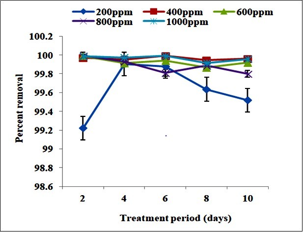
The residual concentrations of lead after treatment with B. subtilis of different preparations were determined (Table 2). For each treatment period, live cells showed the highest value for the residual concentration of lead followed by autoclaved cells and immobilized cells. The biomass (g/ml) of B. subtilis during lead treatment showed that for each day the highest biomass obtained is with 1000 ppm concentration of lead with the maximum after 6days of treatment. It indicates that lead concentration and biomass are directly proportional with each other. Highest biomass is obtained for all the lead concentrations on the sixth day with respect to the treatment period (Figure 2).
Table 2. Residual concentration of lead (ppm) after treatment with Bacillus subtilis of different preparations for 1000 ppm lead concentration| Treatment period | Cell types | ||
| (minutes) | Live | Autoclaved | Immobilized |
| 30 | 44.621 | 12.871 | 0.374 |
| 60 | 51.911 | 27.417 | 14.616 |
| 90 | 40.494 | 13.619 | 8.718 |
| 120 | 20.989 | 19.471 | 12.852 |
| 150 | 28.100 | 14.732 | 11.959 |
Figure 2.Biomass (g/ml) of Bacillus subtilis during lead treatment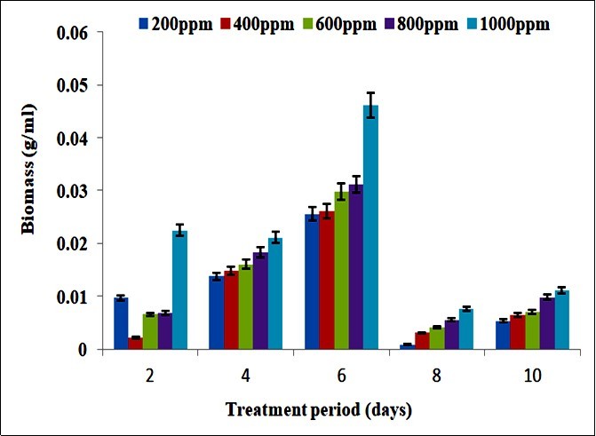
Figure 3 reveals the percent removal of lead after treatment with B. subtilis of different preparations. It infers that immobilized cells are highly efficient in the sorption of lead, while the activity of autoclaved cells is lesser and least for live cells, for all the cases of treatment period. The maximum removal of lead by immobilized cells is observed after 30 mins.
Figure 3.Percent removal of lead after treatment with Bacillus subtilis of different preparations
The influence of various sugars at 10% concentration on the biomass of B. subtilis during lead treatment is exhibited in Figure 4. It shows that biomass is the highest in the case of dextrose, followed by glucose, fructose, sucrose and lactose. This trend indicates that increased biomass is due to the influence of monosaccharides (dextrose, glucose and fructose) while with disaccharides (sucrose and lactose) the biomass decreases.
Figure 4.Influence of sugars on the biomass (g/ml) of Bacillus subtilis during lead treatment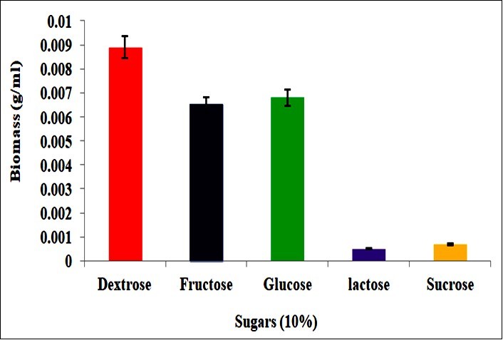
Two way analysis of variance for the factor, residual concentration of lead with the variables, treatment period and lead concentration indicated that the variations in residual concentration of lead due to treatment period and lead concentration were not statistically significant at 5% level. The variations in residual concentration of lead due to cell types were not significant at 5% level but significant due to treatment period. The variations in the biomass (g/ml) of B. subtilis due to lead concentration and treatment period were statistically significant at 5% level.
The variation in percent removal of lead due to cell types was not statistically significant at 5% level but significant due to treatment period. The variation in percent removal of lead due to lead concentration was not statistically significant at 5% level but significant due to treatment period (Table 3).
Table 3. Results of two way analysis of variance (ANOVA) for the various factors during the bioremediation of lead by Bacillus subtilis| Factor | Source of variation | Calculated F value | F table value at 5% level | Level ofsignificance |
| Residual concentration of lead | lead concentration | 0.84 | 3 | Not significant (P>0.05) |
| treatment period | 2.51 | 3 | Not significant (P>0.05) | |
| Residual concentration of lead | cell types | 0.95 | 3.83 | Not significant (P>0.05) |
| treatment period | 11.27 | 4.45 | Significant(P<0.05) | |
| Biomass(g/ml) of B. subtilis | lead concentration | 48.8 | 3 | Significant(P<0.05) |
| treatment period | 8.46 | 3 | Significant(P<0.05) | |
| Percent removal of lead | lead concentration | 0.66 | 3 | Not significant (P>0.05) |
| treatment period | 5.09 | 3 | Significant(P<0.05) | |
| Percent removal of lead | cell types | 0.95 | 3.83 | Not significant (P>0.05) |
| treatment period | 11.27 | 4.45 | Significant(P<0.05) |
The Freundlich adsorption isotherms for lead biosorption by B. subtilis after every two days of treatment period are given in Figure 5. The Langmuir adsorption isotherms for lead biosorption by B. subtilis after every two days of treatment period are given in Figure 6. In Freundlich isotherm models, R2 was the maximum after two days of treatment and it showed a decline with the increase in treatment period. Kf was the highest after eight days of treatment while 1/n was the maximum after four days of treatment. In the case of Langmuir models, R2 and Qmax were the highest after two days of treatment while b was the maximum after eight days of treatment (Table 4).
Figure 5.Freundlich isotherm for lead biosorption by Bacillus subtilis after different days of treatment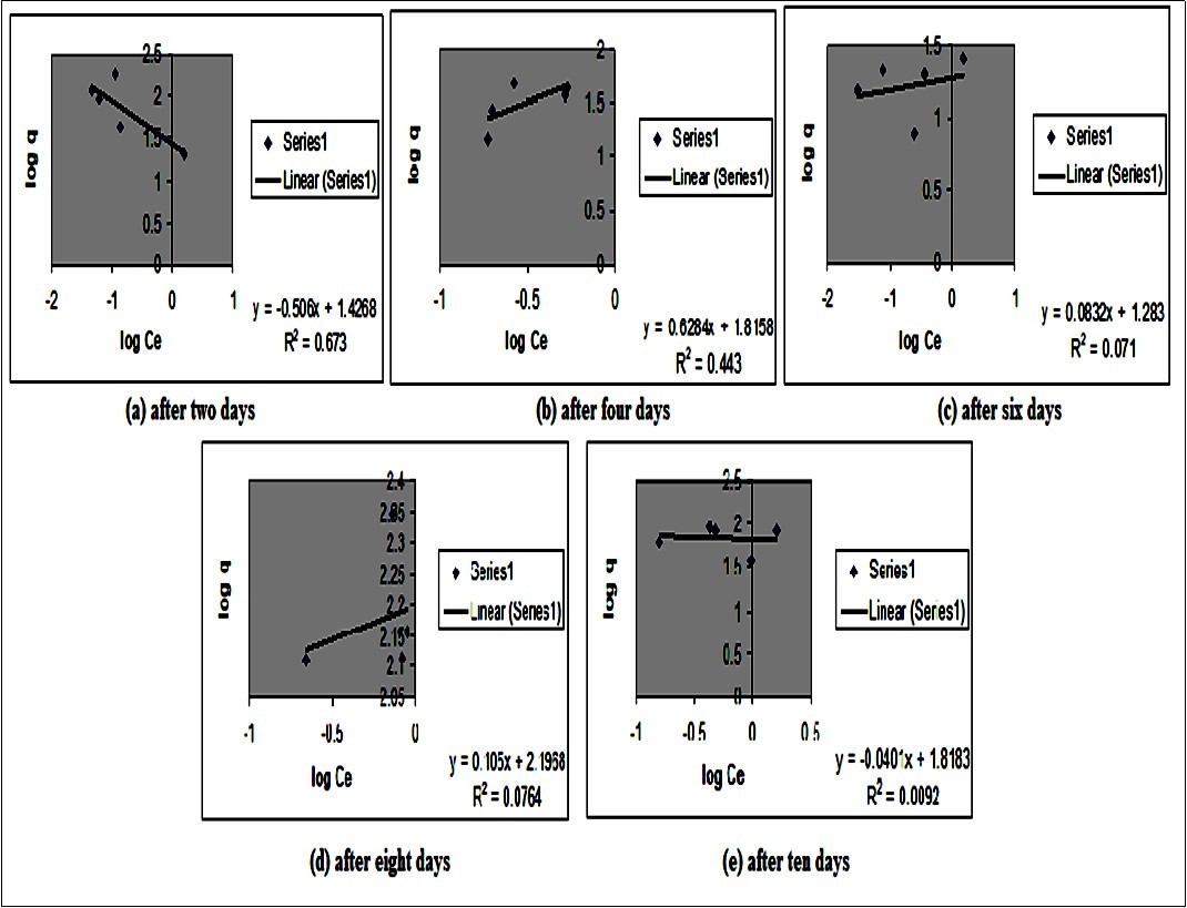
Figure 6.Langmuir isotherm for lead biosorption by Bacillus subtilis after different days of treatment
| Treatment Period (days) | Isotherm constants | ||
| 2 | Freundlich | R2 | 0.673 |
| Kf | 17.78 | ||
| 1/n | -0.445 | ||
| Langmuir | R2 | 0.588 | |
| Qmax (mg/g) | 1000 | ||
| b (L/mg) | -0.001 | ||
| 4 | Freundlich | R2 | 0.443 |
| Kf | 25.12 | ||
| 1/n | 0.364 | ||
| Langmuir | R2 | 0.510 | |
| Qmax (mg/g) | 50 | ||
| b (L/mg) | 0.035 | ||
| 6 | Freundlich | R2 | 0.071 |
| Kf | 1.413 | ||
| 1/n | 0.176 | ||
| Langmuir | R2 | 0.001 | |
| Qmax (mg/g) | 16.66 | ||
| b (L/mg) | -3.450 | ||
| 8 | Freundlich | R2 | 0.076 |
| Kf | 133.4 | ||
| 1/n | 0.287 | ||
| Langmuir | R2 | 0.117 | |
| Qmax (mg/g) | 161.29 | ||
| b (L/mg) | 0.044 | ||
| 10 | Freundlich | R2 | 0.009 |
| Kf | 5.623 | ||
| 1/n | -0.017 | ||
| Langmuir | R2 | 0.009 | |
| Qmax (mg/g) | 71.42 | ||
| b (L/mg) | 0.079 | ||
Discussion
Even low levels of lead can cause permanent damage in organisms. The immediate measure to prevent lead poisoning is to avoid exposure to lead. Removal of the source of lead is critical to reducing lead levels. Biosorption is an alternative to traditional physico-chemical means for removing toxic metals from ground waters and waste waters. Removal of lead from solution was studied using growing cells and washed cells of Bacillus cereus 29. The ability of the strain, B. subtilis to remove lead from solution was investigated in the present study. B. subtilis was reported to be the best for biosorbing lead among copper, zinc, lead and cadmium 30. B. subtilis, as a Gram positive organism has high capacity towards the binding with heavy metals, when compared with Gram negative bacteria because of their different cell wall structures. Phosphate groups present in the teichoic acids and other associated acids present in the Gram positive cell walls are the key factors responsible for the uptake of heavy metals present in the environment. The most important thing in the uptake of heavy metals are carboxyl groups, the sources which are the teichoic acids in connection with peptidoglycon layers present in cell wall 31, 32, 33.
When different concentrations of lead were tested, the strain B. subtilis was able to resist upto 1000 ppm. When the walls of B. subtilis were tested for the uptake of eighteen different metals, lead was shown to be taken up in small amounts into the wall 34. Maximum percent removal of lead was observed at 1000 ppm and minimum at 100 ppm. It indicates that at higher concentrations of lead, the uptake by B.subtilisis efficient. Efficient uptake of lead by B. subtilis can be explained on the basis of presence of some metal binding moieties on the surface of B. subtilis. The important role of negatively charged carboxyl groups in lead biosorption by acetone washed biomass of Saccharomyces uvarum was demonstrated 35.
With different concentrations of lead, the biomass (g/ml) of B.subtilis obtained was the highest at 1000 ppm. It shows that biomass is directly proportional to increase in lead concentration. However, after six days the biomass again showed a decline. It indicates the saturation of metal binding moieties of B.subtilis. Lead ions show more affinity to algal biomass and they bind through a combination of ion-exchange, chelation and reduction reactions, along with metallic lead precipitation on the cell wall 36. When different preparations (live, dead and immobilized cells) of B. subtilis were exposed to 1000 ppm concentration of lead, immobilized cells, showed the highest percent removal. The highest biosorption of lead among Cu, Cd and Pb with multiple fixed beads containing immobilized bacterial biomass was studied 37. Two immobilized marine algae Sargassum fluitans and Ascophyllum nodosum were used for the uptake of Cd, Cu, Ni, Pb and Zn. Both algae showed highest uptake for Pb 38. In the present study, autoclaved (dead) cells showed a higher percentage of lead removal than normal cells. In contrast with live cells, dead cells have several merits such as the capacity to treat huge amount of wastewater with less concentration of metals, less operation time, accessible without any dangerous byproducts and the lack of constraints on enzymatic activities produced by metal adsorption 39. Along with that, the dead cells do not need a constant nutrients requirement and are not influenced by toxic wastes. It can be redeveloped and reused for several cycles. Therefore, the application of dead microbial cells in this practice has added benefits in removal of heavy metals from waste water 40.
In the present study, supplementation of different sugars (Dextrose, fructose, glucose, lactose and sucrose) indicated that dextrose, fructose and glucose were efficiently utilized by B. subtilis, which enhanced the biomass. The biomass was higher in the case of monosaccharides (dextrose, glucose and fructose) than that of disaccharides (lactose and sucrose) which imply that due to simple configuration of monosaccharides they are easily taken up by B. subtilis than disaccharides. Effect of carbon sources on dye decolourization by Aspergillus sp. was studied and it was demonstrated that the fungus reduces colour of dye by 98% in the presence of glucose (monosaccharide) and 92% in presence of sucrose (disaccharide) 41.
The biosorption isotherm curve denotes the equilibrium distribution of metal ions between the aqueous and solid phases. The equilibrium distribution is very essential in finding out the highest biosorption potential. Various isotherm models are used to portray this equilibrium distribution. Langmuir and Freundlich models are extensively used in equilibrium analysis to analyse the mechanism of sorption 42, 43, 44, 45. The Langmuir model considers sorption by monolayer type and presumes that all sorbent surface active sites have the similar affinity towards heavy metal ions 46. The Freundlich isotherm is an experimental equation which assumes a various biosorption system with diverse active sites 47. In the present study, the Freundlich and Langmuir isotherm coefficients were R2= 0.673, Kf= 17.78, 1/n= -0.4452, R2= 0.5884, Qmax= 1000 and b= -0.0014. Ray et al. (2005) studied the bioaccumulation of Pb (II) by Bacillus cereus (14). The isotherm coefficients were R2= 0.988, Kf= 13.487, 1/n= 0.47, R2= 0.941, Qmax= 70.423 and b= 0.214. Hence, it can be concluded that uptake of lead increased with increased concentration of lead. Biomass is also observed to be influenced by increasing lead concentration. Immobilized cells are shown to be highly efficient in lead uptake. Monosaccharides are observed to be effective on the biomass (g/ml) of B.subtilis.
Conclusion
Bioremediation is the major reason for the disappearance of these heavy metals from contaminated sites. However, the process is influenced by various environmental conditions like pH and temperature. The studies so far carried out suggest that B. subtilis endowed with all this properties of bioremediation enabled the biosorption and bioaccumulation of lead. Hence B.subtilis can be employed as an efficient organism for the effective uptake of lead and other heavy metals.
Acknowledgments
The authors thank the authorities of the American College, Madurai for the facilities and Tamil Nadu State Council for Science and Technology (TNSCST) Chennai for the financial assistance.
References
- 1.Sardar K, Ali S, Hameed S, Afzal S, H M Tauqeer. (2013) Heavy metals contamination and what are the impacts on living organisms”. , Greener J.Envt.Magt. Pub. Safety 2, 172-179.
- 2.Mahmood Q, Rashid A, S, M R Azim, Bilal M. (2012) Current status of toxic metals addition to environment and its consequences”. In: The plant family Brassicaceae.Springer. , Dordrecht 35-69.
- 3.Gupta R, Mohapatra H. (2003) Microbial biomass: An economical alternative for removal of heavy metals from waste water”.IndianJ.Exp.Biol. 41, 945-966.
- 4.Jaishankar M, Tseten T, Anbalagan N, B, K N Beeregowda. (2014) Toxicity, mechanism and health effects of some heavy metals”. , Interdisciplinary toxicology 7(2), 60-72.
- 5.G M Naja, Volesky B. (2009) Toxicity and sources of Pb, Cd, Hg, Cr, As, and radionuclides in the environment”. Heavy metals in the environment. 8, 16-18.
- 6.Morais S, F G Costa, Pereira M D L. (2012) Heavy metals and human health”. Environmental health–emerging issues and practice. 10, 227-246.
- 7.Tong S, Schirnding Y E V, Prapamontol T. (2000) Environmental lead exposure: a public health problem of global dimensions”. , Bulletin of the world health organization 78, 1068-1077.
- 8.Sardar K, Ali S, Hameed S, Afzal S, Fatima S et al. (2013) Heavy metals contamination and what are the impacts on living organisms”. , Greener Journal of Environmental management and public safety 2(4), 172-179.
- 9.Flora G, Gupta D, Tiwari A. (2012) Toxicity of lead: a review with recent updates”. , Interdisciplinary toxicology 5(2), 47-58.
- 10.Crini G, Lichtfouse E. (2019) Advantages and disadvantages of techniques used for wastewater treatment”. , Environmental Chemistry Letters 17(1), 145-155.
- 11.G M Naja, Volesky B. (2010) Treatment of metal-bearing effluents: removal and recovery”. Handbook on heavy metals in the environment. Taylor & Francis. , Boca Raton 247-291.
- 12.A K Verma, R, Bhunia P. (2012) A review on chemical coagulation/flocculation technologies for removal of colour from textile wastewaters”. , Journal of environmental management 93(1), 154-168.
- 13.Gaur N, Flora G, Yadav M, Tiwari A. (2014) A review with recent advancements on bioremediation-based abolition of heavy metals”. Environmental Science: Processes & Impacts 16(2), 180-193.
- 14.Gavrilescu M. (2004) Removal of heavy metals from the environment by biosorption”. , Engineering in Life Sciences 4(3), 219-232.
- 15.Juwarkar A A, Singh S K, Mudhoo A. (2010) A comprehensive overview of elements in bioremediation”. Reviews in Environmental Science and bio/technology 9(3), 215-288.
- 16.Kumar A, Bisht B S, V D Joshi, Dhewa T. (2011) Review on bioremediation of polluted environment: a management tool”. International journal of environmental sciences. 1(6), 1079-1093.
- 17.Abdi O, Kazemi M. (2015) A review study of biosorption of heavy metals and comparison between different biosorbents”. , J Mater Environ Sci 6(5), 1386-1399.
- 18.Das N. (2010) Recovery of precious metals through biosorption— a review”. Hydrometallurgy,103(1-4) 180-189.
- 19.Fu F, Wang Q. (2011) Removal of heavy metal ions from wastewaters: a review”. , Journal of environmental management 92(3), 407-418.
- 20.Sher S, Rehman A. (2019) Use of heavy metals resistant bacteria—a strategy for arsenic bioremediation”. Applied microbiology and biotechnology. 103(15), 6007-6021.
- 21.Prabhakaran P, M A Ashraf, W S Aqma. (2016) Microbial stress response to heavy metals in the environment’. , RscAdvances 6(111), 109862-109877.
- 22.K E Giller, Witter E, S P Mcgrath. (1998) Toxicity of heavy metals to microorganisms and microbial processes in agricultural soils: a review”. Soil biology and biochemistry,30(10-11). 1389-1414.
- 23.Silva Da, B S, V, Ayub M A Z. (2014) Production and optimization of poly-γ-glutamic acid by Bacillus subtilis BL53 isolated from the Amazonian environment”. Bioprocess and biosystems engineering. 37(3), 469-479.
- 24.C R Harwood. (1992) Bacillus subtilisand its relatives: molecular biological and industrial workhorses”. Trends in biotechnology. 10, 247-256.
- 25.J K Roy, S K Rai, A K Mukherjee. (2012) Characterization and application of a detergent-stable alkaline α-amylase fromBacillus subtilisstrain AS-S01a”. International journal of biological macromolecules. 50(1), 219-229.
- 26.Puvaneswari N, Muthukrishnan J, Gunasekaran P. (2002) Bioremediation of benzidine based azo dyes direct red and direct blue by the immobilized cells ofPseudomonas fluorescensD41”.IndianJ.Exp.Biol. 40, 1131-1136.
- 28.Langmuir I. (1916) The adsorption of gases on plane surface of glass, mica and platinum”. , J. Am. Chem. Soc 40, 1361-1368.
- 29.Ray L, Paul S, Bera D, Chattopadhyay P. (2005) Bioaccumulation of Pb (II) from aqueous solution byBacillus cereus”.J.Hazd..Subst.Res. 5, 1-21.
- 30.Costa A C A, F P Duta. (2001) Bioaccumulation of copper, zinc, cadmium and lead byBacillussp.”.Braz. , J.Microbiol 32, 1-5.
- 31.M U, Kemper M, Doyle R, T J Beveridge. (1992) The membrane-induced proton motive force influences the metal binding ability ofBacillus subtiliscell walls”. , Applied and Environmental Microbiology 58(12), 3837-3844.
- 32.Remade J. (1990) The cell wall and metal binding. Biosorption of heavy metals”. , Boca Raton 83-92.
- 33.Brown S, Santa Maria Jr, P J, Walker S. (2013) Wall teichoic acids of Gram-positive bacteria”. , Annual review of microbiology 67, 313-336.
- 34.T J Beveridge, R G Murray. (1976) Uptake and retention of metals by cell walls ofBacillus subtilis”.J.Bacteriol. 127-1502.
- 35.Ashkenazy R, Gottlieb L, Yannai S. (1997) Characterization of acetone-washed yeast biomass functional groups involved in lead. , Biosorption”.Biotech.Bioengg 55, 1-10.
- 36.Raize O, Argaman Y, Yannai S. (1995) Mechanisms of biosorption of different heavy metals by brown marine macroalgae”.J.Chem.Tech.Biotech. 63, 257-261.
- 37.J S Chang, J C Huang. (1998) Selective adsorption/recovery of Pb, Cu and Cd with multiple fixed beds containing immobilized bacterial biomass”.Biotech. , Prog 14, 735-741.
- 38.Volesky B, Z R Holan, Leusch A. (1995) Biosorption of heavy metals (Cd, Cu, Ni, Pb, Zn) by chemically reinforced biomass of marine algae”.J. , Chem. Tech. Biotech 62, 279-288.
- 39.Hemambika B, M J Rani, V R Kannan.Biosorption of heavy metals by immobilized and dead fungal cells: A comparative assessment.J. , Ecol. Nat. Environ.,2011 3, 168-175.
- 40.Aksu Z.Application of biosorption for the removal of organic pollutants:. , A review.ProcessBiochem.,2005 40, 997-1026.
- 41.Gupta P, Goyal D. (2007) . , Decolourization of synthetic dye byAspergillussp.”.IndianJ.Microbiol 44, 191-195.
- 42.K A Shroff, V K. (2011) Kinetic and equilibrium studies on biosorption of nickel from aqueous solution by dead fungal biomass ofMucorhiemalis.Chem. , Eng 171, 1234-1245.
- 43.R M Gabr, Hassan S H A, Shoreit A A M. (2008) Biosorption of lead and nickel by living and non-living cells ofPseudomonas aeruginosaASU 6a.Int.Biodeter.Biodegr.62. 195-203.
- 44.Joo J H, Hassan SHA, Oh S E. (2010) Comparative study of biosorption of Zn2+byPseudomonas aeruginosaandBacillus cereus”.Int.Biodeter.Biodegr. 64, 734-741.
- 45.Ji Y, Gao H, Sun J, Cai F. (2011) Experimental probation on the binding kinetics and thermodynamics of Au(III) ontoBacillus subtilis”.Chem. , Eng 172, 122-128.
Cited by (3)
- 1.Gana, A. J. , Tijjani, M. B. , Akinyelure, E. O. , 2021, Removal of Lead Ions from Water Using Pellet Generated from Bacillus subtilis Isolated from Gold Mining Site in Niger State, UMYU Journal of Microbiology Research (UJMR), 6(1), 105, 10.47430/ujmr.2161.014
- 2.James Nilina, Umesh Mridul, 2024, Multifarious Potential of Biopolymer-Producing Bacillus subtilis NJ14 for Plant Growth Promotion and Stress Tolerance in Solanum lycopercicum L. and Cicer arietinum L: A Way Toward Sustainable Agriculture, Molecular Biotechnology, 66(5), 1031, 10.1007/s12033-023-01001-9
- 3.Monga Yukti, Sharma Shivangi, Singh Shivendra, Gupta Ashu, 2024, Biochar, Clay, Zeolites, and Microorganism-based Methods for Remediation of Heavy Metals, Current Green Chemistry, 11(1), 2, 10.2174/2213346110666230915140448